Elly K. Riley, DO, FACOFP
This paper was submitted as part of Dr. Riley's application for ACOFP Fellowship, which recognizes exceptional national, state, and local service through teaching, authorship, research, or professional leadership. Visit the ACOFP Fellows page to learn more about fellowship and the nomination process
A fifteen day-old female presented to the outpatient family medicine clinic for a two week well child visit. She was born at 41 weeks via spontaneous vaginal delivery, to a fifteen-year-old G1 without complications during the delivery or birth. Birth weight was seven pounds, zero ounces.
On day of exam her weight was seven pounds, 9 ounces (26th percentile). Exam was notable for a four and a half centimeter comma-shaped plaque with abnormal hair growth and mildly hypertrophic edges with minimal diffuse erythema on the left temporo-parietal scalp. Fine, short hair growth was noted across the lesion.

Figure 1. scalp lesion, 15 day old female
QUESTIONS
- What is the diagnosis based on the case and clinical image?
a. Epidermal nevus
b. Nevus sebaceous
c. Nevus psiloliparus
d. Aplasia cutis congenital
2. What is the most common serious complication of this condition?
a. Developmental delay
b. Epilepsy
c. Basal cell carcinoma
d. There are no serious complications of this disorder
3. What is the preferred long term management of this condition?
a. Close monitoring with excision for concerning clinical changes
b. Excision
c. Excision with skin grafting
d. Lymphatic OMT
ANSWERS
- What is the diagnosis based on the case and clinical image?
Correct answer:
b. Nevus sebaceous
- What is the most common serious complication of this condition?
Correct answer:
c. basal cell carcinoma
- What is the preferred long term management of this condition?
Correct answer:
a. Close monitoring with excision for concerning clinical changes
CLINICAL COURSE OF PATIENT AND DISCUSSION
The patient’s mother was advised that she will eventually be referred to dermatology as she has a small risk of developing a benign or malignant tumor within the nevus sebaceous itself. They returned six months later; mother had concerns about the skin lesion, specifically that she thought the skin lesion became very red when the patient cried, and concerned that it might affect her underlying brain development. Parents were reassured, but referred to dermatology, and advised to follow up for annual well child exam and monitoring.
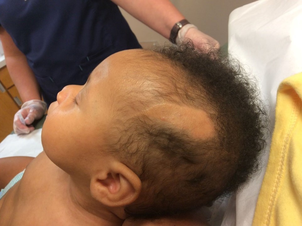
Figure 2. Nevus sebaceous at 6 months of age.
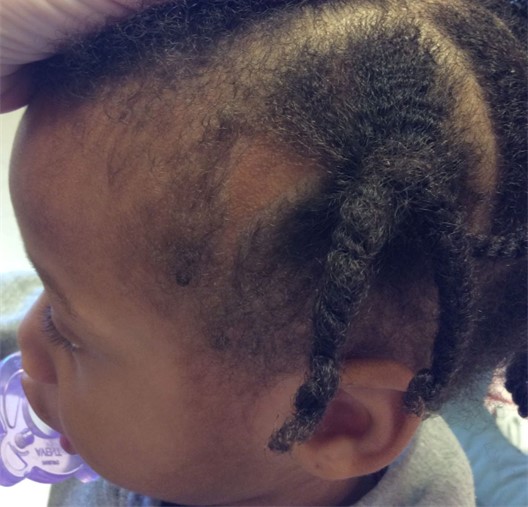
Figure 3. Nevus sebaceous at 35 months of age, partially obscured by hairstyle.
NEVUS SEBACEOUS
Nevus sebaceous, sometimes referred to as nevus sebaceous of Jadassohn is an uncommon, benign tumor of the skin, occurring in 0.3 percent of newborns (3,4). They are typically present at birth, containing immature hair follicles, and represent an overgrowth of the sebaceous gland. These congenital skin lesions may present as a linear, ovoid, or round plaque, waxy in appearance, and are typically salmon, tan or yellow in color. (2). Nevus sebaceous is most frequently found on the scalp, unlike epidermal nevi, which are typically located on the trunk and limbs, and less commonly on the scalp or face (4). Nevus sebaceous, however, may occur on other parts of the body. The lesion typically becomes more hypertrophic with age, and has a 10-30% incidence of developing a benign or malignant neoplasm within the primary lesion itself (3). Although nevus sebaceous is a clinical diagnosis, some clinicians may want to confirm the diagnosis with biopsy. However, nevus sebaceous has unique characteristics that may allow experienced physicians to utilize ultrasound to help confirm the diagnosis, eliminating the need for biopsy and painful, potentially traumatic procedures on what is frequently a pediatric patient. (1) Ultrasound also has the potential to be used as a tool for monitoring the lesion over time. Differential diagnosis for nevus sebaceous includes epidermal nevus, nevus psiloliparis, aplasia cutis congenital, solitary mastocytoma, juvenile xanthogranuloma, and syringocystadenoma papilliferum (4).
Nevus Sebaceous pathophysiology
Nevus sebaceous originates from both epithelial and nonepithelial cell lines and is an example of how the osteopathic tenet that structure and function are reciprocally interrelated can go awry during the embryologic period. The natural progression of nevus sebaceous lesions typically evolves through three overlapping stages. The first stage, during the infant years, is characteristic of papillomatous hyperplasia and fine irregular hair growth stemming from immature and abnormally formed hair follicles. Small sebaceous glands may also be present. The second stage involves nevus expansion during puberty, when sebaceous and apocrine glands all over the body are rapidly developing. Sebaceous glands in the NS lesion are abnormally located in high numbers in the dermis. During this stage, the nevus typically evolves from a flat plaque to a more verrucous plaque. The last stage involves the development of benign and malignant epithelial neoplasms (1,2).
Tumors most commonly arising from nevus sebaceous lesions include basal cell carcinomas (the most commonly reported malignant tumor), trichoblastomas (the most commonly reported benign tumor), sebaceous carcinomas, and squamous cell carcinomas (2).
Nevus Sebaceous Management
Because of the risk of developing a malignant epithelial neoplasm, full thickness excision of both epidermis and dermis was historically suggested as primary management and prophylaxis, although debate was ongoing as to if and when excision was necessary. Most tumors develop during the third stage of NS, during adulthood, but can also occur during childhood, not excluding malignancies. More contemporary recommendations are to evaluate each case individually to determine need for excision (5). Referral to dermatology or plastic surgery may be necessary; at minimum, annual examination and parent education as to changes to watch out for are key to appropriate timing of excision, should clinical signs of neoplasms develop.
CONCLUSION
Nevus sebaceous is an uncommon, but important diagnosis given its presence at birth, and risk of developing a malignancy later in life. The osteopathic family physician is prepared by their education and training to perform a thorough exam, and should be prepared to list nevus sebaceous in the differential diagnosis of an undifferentiated congenital plaque, coordinating appropriate care for the patient on a case by case basis.
REFERENCES
- Wortsman X, Ferreira-Wortsman C, Corredoira Y. Ultrasound Imaging of Nevus Sebaceous of Jadassohn. J Ultrasound Med. 2021 Feb;40(2):407-415. doi: 10.1002/jum.15405. Epub 2020 Jul 29. PMID: 32725836.
- Moody MN, Landau JM, Goldberg LH. Nevus sebaceous revisited. Pediatr Dermatol. 2012 Jan-Feb;29(1):15-23. doi: 10.1111/j.1525-1470.2011.01562.x. Epub 2011 Oct 13. PMID: 21995782.
- Kakagia DD, Christodoulou KC, Noulas CN, Stouras I, Fiska A. A Linear Waxy Verrucous Plaque of the Scalp. Cureus. 2023 Apr 20;15(4):e37881. doi: 10.7759/cureus.37881. PMID: 37223135; PMCID: PMC10202673.
- Wright, TS (2020). Nevus sebaceus and nevus sebaceus syndrome. In M.L. Levy, & R. Corona (Eds.), UpToDate. Available from https://www.uptodate.com/contents/nevus-sebaceus-and-nevus-sebaceus-syndromes (Accessed on March 5, 2020)
- Patel P, Malik K, Khachemoune A. Sebaceus and Becker's Nevus: Overview of Their Presentation, Pathogenesis, Associations, and Treatment. Am J Clin Dermatol. 2015 Jun;16(3):197-204. doi: 10.1007/s40257-015-0123-y. PMID: 25782676.